
Skin cancer is the most common cancer in the United States, affecting millions of people each year. Fortunately, when detected early, many skin cancers can be treated successfully. Regular skin cancer screenings play an important role in identifying suspicious lesions before they become more advanced.
At Rangel Dermatology, we provide comprehensive skin cancer screenings in Miami for patients of all ages. Whether you have a personal history of skin cancer, a family history of melanoma, numerous moles, or simply want a professional skin examination, regular screenings can help detect skin cancer at its earliest and most treatable stages.
Why Is Skin Cancer So Common in Miami?
Living in Miami and South Florida means enjoying sunshine year-round, but it also means increased exposure to ultraviolet (UV) radiation.
Ultraviolet radiation from the sun is one of the primary causes of skin cancer. Over time, cumulative sun exposure can damage the DNA within skin cells, leading to the development of cancerous growths.
Many South Florida residents spend significant time outdoors participating in activities such as boating, beach outings, golf, tennis, pickleball, running, and other outdoor sports. As a result, skin cancer is a common condition that dermatologists diagnose and treat throughout the Miami area.
Because UV damage accumulates over a lifetime, even sun exposure that occurred years or decades ago can contribute to the development of skin cancer later in life.
What Types of Skin Cancer and Precancerous Lesions Are We Looking For?
During a skin cancer screening, we carefully evaluate the skin for precancerous lesions, common skin cancers, and less common skin malignancies.
Basal Cell Carcinoma
Basal cell carcinoma is the most common type of skin cancer. It often appears as a pearly bump, non-healing sore, pink patch, or scar-like area on sun-exposed skin.
Although basal cell carcinoma rarely spreads to other parts of the body, it can continue to grow locally and cause significant tissue destruction if left untreated.
Squamous Cell Carcinoma
Squamous cell carcinoma is the second most common type of skin cancer. It may present as a scaly patch, rough bump, non-healing sore, or tender growth.
Unlike basal cell carcinoma, squamous cell carcinoma has a greater potential to spread if not diagnosed and treated promptly.
Actinic Keratoses (Precancerous Lesions)
During a skin cancer screening, we also look for actinic keratoses, commonly called AKs. These are rough, scaly spots caused by cumulative sun damage and are considered precancerous lesions.
Actinic keratoses most commonly develop on sun-exposed areas such as the face, scalp, ears, chest, forearms, and hands. They may feel rough like sandpaper and can sometimes be easier to feel than see.
While not all actinic keratoses progress to skin cancer, they have the potential to develop into squamous cell carcinoma over time. For this reason, dermatologists often recommend treatment when these lesions are identified.
Common treatment options include liquid nitrogen cryotherapy (freezing), prescription topical medications, photodynamic therapy (PDT), and other field-directed treatments for extensive sun damage.
Identifying and treating actinic keratoses is an important part of skin cancer prevention and is a routine component of comprehensive skin cancer screenings.
Melanoma
Melanoma is the most serious form of skin cancer because of its ability to spread to other organs.
Melanoma can develop within an existing mole or arise as a new dark spot on the skin. Early detection is critical, as treatment is most successful when melanoma is identified in its earliest stages.
The ABCDEs of Melanoma
One of the ways dermatologists evaluate moles is by looking for the ABCDE warning signs of melanoma. While not every melanoma follows these rules, the ABCDEs provide a helpful framework for identifying potentially concerning lesions.
A – Asymmetry
One half of the mole does not match the other half.
B – Border Irregularity
The edges appear uneven, scalloped, blurred, or poorly defined.
C – Color Variation
Multiple colors within the same lesion, such as shades of brown, black, tan, red, white, or blue.
D – Diameter
A lesion larger than 6 millimeters (about the size of a pencil eraser) may warrant evaluation, although melanomas can be smaller.
E – Evolving
Any mole or spot that is changing in size, shape, color, elevation, or symptoms such as itching, bleeding, or crusting should be examined by a dermatologist.
In many cases, the “E” for evolving is the most important warning sign. A mole that looks different from your other moles or is changing over time deserves prompt evaluation.
Other Types of Skin Cancer
Although less common, skin cancer screenings also evaluate for other malignancies, including:
- Merkel cell carcinoma
- Dermatofibrosarcoma protuberans (DFSP)
- Sebaceous carcinoma
- Cutaneous lymphoma
- Other rare skin tumors
Board-certified dermatologists are trained to recognize both common and uncommon skin cancers, many of which can mimic benign skin growths.
What Happens During a Skin Cancer Screening?
A skin cancer screening is a thorough examination of the skin performed by a board-certified dermatologist.
When you arrive for your appointment, you will be asked to remove your clothing except for your underwear and change into a paper gown.
Dr. Rangel will then perform a comprehensive examination of your skin from head to toe, looking for suspicious moles, growths, or other lesions that could represent skin cancer or precancerous changes.
Because skin cancer can occur anywhere on the body, including areas that receive little sun exposure, patients may be asked to adjust their underwear or gown as needed so these areas can be properly examined.
The examination is painless and typically takes only a few minutes.
What Is a Dermatoscope?
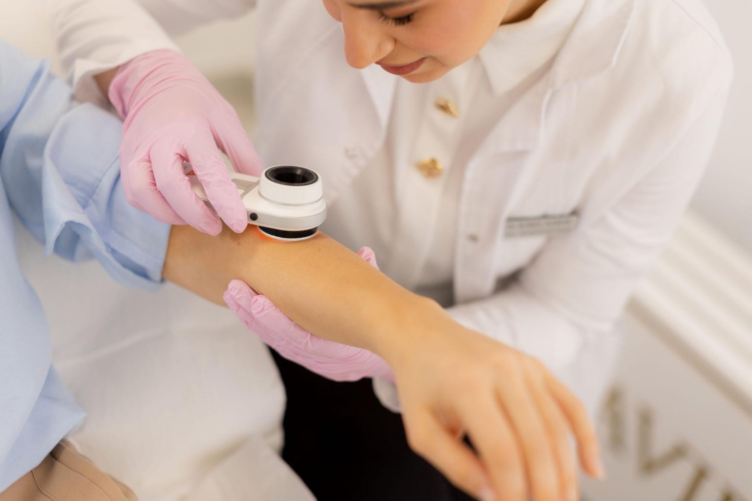
One of the most important tools used during a skin cancer screening is a dermatoscope.
A dermatoscope is a specialized handheld device that allows dermatologists to examine structures beneath the surface of the skin that are not visible to the naked eye.
Using dermoscopy, Dr. Rangel can evaluate:
- Pigment patterns within moles
- Blood vessel patterns
- Colors and structures within skin lesions
- Features that may suggest skin cancer
Dermoscopy significantly improves the accuracy of skin cancer detection and helps distinguish benign lesions from potentially concerning growths.
What Happens If Something Looks Suspicious?
If a lesion appears suspicious for skin cancer, a biopsy is often recommended.
A skin biopsy is a simple in-office procedure in which a small sample of tissue is removed and sent to a dermatopathology laboratory for microscopic evaluation.
The biopsy allows us to determine whether a lesion is benign, precancerous, or cancerous and helps guide the next steps in treatment.
Depending on the diagnosis, treatment may include:
- Surgical excision
- Mohs surgery
- Additional skin cancer surveillance
- Other specialized treatments
If skin cancer is detected, we will review the pathology results with you and develop an individualized treatment plan.
How Often Should You Have a Skin Cancer Screening?
For many patients without a personal history of skin cancer, an annual skin cancer screening is recommended.
However, some patients may benefit from more frequent examinations, including those with:
- A personal history of skin cancer
- A family history of melanoma
- Numerous atypical moles
- Significant sun damage
- A weakened immune system
- Prior organ transplantation
Patients with a history of skin cancer often require screenings every three to six months, depending on their individual risk factors and cancer history.
When Should You See a Dermatologist?
You should schedule a skin cancer screening if you:
- Have never had a professional skin examination
- Notice a new or changing mole
- Notice a spot that meets any of the ABCDE criteria for melanoma
- Have a personal or family history of skin cancer
- Have significant sun exposure or sun damage
- Have a lesion that bleeds, crusts, or does not heal
Early detection remains one of the most effective tools for reducing the impact of skin cancer.
Skin Cancer Screening at Rangel Dermatology
At Rangel Dermatology, Dr. Lauren Rangel provides comprehensive skin cancer screenings in Miami using detailed clinical examinations and dermoscopy to evaluate moles, growths, and other skin lesions.
Whether you are due for your annual skin check or have concerns about a changing spot, we are committed to providing thorough, personalized care focused on early detection and skin cancer prevention.
If you are looking for a board-certified dermatologist in Miami for a skin cancer screening, contact Rangel Dermatology to schedule your appointment.